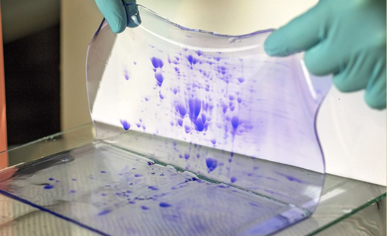
Phosphorylated proteins play a pivotal role in a multitude of cellular processes, acting as molecular switches that regulate key functions within the cell. Understanding the phosphorylation status of proteins is crucial for unraveling the mysteries of cell signaling pathways, diseases, and drug development. In this blog post, provided by Kendrick Labs, Inc, we delve into the significance of Western blot in the analysis of phosphorylated proteins, exploring its principles, techniques, and best practices.
The Basics of Phosphorylation
Before we delve into the world of Western blotting, it’s essential to grasp the concept of phosphorylation. Phosphorylation is a post-translational modification that involves the addition of a phosphate group to specific amino acids in a protein. This modification is a fundamental regulatory mechanism in biology, and it controls a vast array of cellular processes, including cell growth, differentiation, and response to external stimuli.
Phosphorylation events are dynamic and reversible, making them pivotal in cell signaling pathways. Dysregulation of phosphorylation can lead to various diseases, including cancer, diabetes, and neurodegenerative disorders. Consequently, understanding phosphorylation is essential for both basic research and clinical applications.
The Role of Western Blot in Phosphorylated Protein Analysis
Western blotting, also known as immunoblotting, is a widely used technique for the detection and quantification of specific proteins within a sample. It has proven to be indispensable in phosphorylated protein research due to its sensitivity and specificity.
1. Antibody-Based Detection
At the core of Western blotting is the use of specific antibodies that can recognize and bind to the protein of interest, including phosphorylated proteins. These antibodies are often designed to target the phosphorylated amino acids or motifs unique to the phosphorylated state. The specificity of these antibodies is what makes Western blot an invaluable tool in phosphorylated protein analysis.
2. Separation by Gel Electrophoresis
Before Western blotting can take place, proteins in a sample must be separated based on their molecular weight. This is achieved through gel electrophoresis, a technique that separates proteins in a sample by size. For phosphorylated protein research, polyacrylamide gels are often used to achieve the required resolution.
3. Transfer to Membrane
Once proteins are separated, they need to be transferred from the gel onto a membrane. This transfer process allows the proteins to be probed with specific antibodies and facilitates subsequent detection.
4. Antibody Binding and Visualization
Following the transfer, the membrane is exposed to antibodies that recognize the phosphorylated protein of interest. This binding event is then visualized using various techniques, such as chemiluminescence or fluorescent detection.
5. Quantification and Analysis
Western blotting provides not only qualitative but also quantitative data on the levels of phosphorylated proteins in a sample. This data is crucial for understanding the dynamics of phosphorylation events and comparing samples under different experimental conditions.
Tips for Successful Western Blot Phosphorylated Protein Analysis
To ensure the success of your Western blot experiments in phosphorylated protein research, it’s essential to follow best practices. Here are some tips to help you achieve accurate and reliable results:
1. Sample Preparation
Proper sample preparation is crucial. Ensure your samples are treated appropriately, including protein extraction and denaturation. Be mindful of preserving the phosphorylation state during sample handling.
2. Gel Electrophoresis
Choose the appropriate gel percentage and running conditions to separate phosphorylated proteins effectively. Stacking and resolving gels can be tailored to your specific research needs.
3. Transfer Efficiency
Optimize the transfer of proteins from the gel to the membrane to avoid loss of phosphorylated proteins. Use the right transfer buffer, appropriate voltage, and transfer duration.
4. Antibody Selection
Select highly specific antibodies for your phosphorylated proteins of interest. Ensure the antibodies are validated and work well in the context of Western blotting.
5. Blocking and Antibody Incubation
Effective blocking of the membrane helps reduce non-specific binding. Follow recommended blocking protocols and incubate with primary and secondary antibodies under optimal conditions.
6. Detection and Imaging
Choose the right detection method based on the antibodies used. Pay close attention to image exposure and ensure that bands are within the linear range for quantification.
Advanced Western Blot Techniques for Phosphorylated Protein Analysis
As technology advances, researchers have developed more advanced Western blot techniques to enhance the detection and analysis of phosphorylated proteins.
1. Phospho-Specific Antibodies
Phospho-specific antibodies are designed to recognize a particular phosphorylated amino acid within a protein. They provide a higher level of specificity and are valuable tools for assessing the phosphorylation state of specific residues.
2. Multiplexing
Multiplexing allows the detection of multiple phosphorylated proteins in a single blot, saving time and reducing sample consumption. This technique is particularly useful when studying complex signaling pathways.
3. Phospho-IP Western Blot
Combining immunoprecipitation (IP) with Western blotting enables the isolation and analysis of phosphorylated proteins in complex mixtures. This approach is highly effective in studying protein-protein interactions in signaling pathways.
4. Phospho-kinase Arrays
Phospho-kinase arrays are valuable tools for simultaneously assessing the phosphorylation status of multiple kinases. These arrays are instrumental in profiling kinase activity in various experimental conditions.
Applications of Western Blot Phosphorylated Protein Analysis
The application of Western blot in phosphorylated protein analysis is broad and impacts various fields of research and clinical diagnostics.
1. Cancer Research
Understanding the phosphorylation status of proteins in cancer cells is critical for identifying potential therapeutic targets and biomarkers. Western blotting plays a significant role in uncovering aberrant signaling pathways in cancer.
2. Drug Development
Phosphorylated proteins are often the targets of drug development efforts. Western blotting is used to validate the effectiveness of drugs in modulating specific phosphorylation events.
3. Signal Transduction Studies
In cell signaling studies, Western blotting helps elucidate the intricate details of phosphorylation events, allowing researchers to map signaling pathways and identify key components.
4. Disease Biomarker Discovery
Phosphorylated proteins can serve as biomarkers for various diseases. Western blot analysis helps identify and validate these biomarkers for diagnostic and prognostic purposes.
Call to Action
As you dive deeper into the world of phosphorylated protein research, mastering the art of Western blotting is paramount. Kendrick Labs, Inc is here to support your journey with cutting-edge products, expertise, and resources. Explore our range of high-quality antibodies, reagents, and instruments designed to enhance your Western blotting experiments.
Whether you’re a seasoned researcher or just beginning your exploration of phosphorylated proteins, we’re your partner in unraveling the secrets of cellular signaling. Visit Kendrick Labs, Inc today to discover the tools you need for successful Western blot experiments and pave the way for groundbreaking discoveries in phosphorylated protein research.
Unlock the potential of Western blotting and stay at the forefront of scientific innovation. Begin your journey with Kendrick Labs, Inc.


